| |
Contents | EBJ Home | Single molecule measurements and biological motors |
There are a number of different ways of adapting a microscope to perform TIRF; the method chosen depends on a number of factors, including the design of microscope, the form of illumination, the specimen and the type of experiment to be performed. For a good general introduction (with a cell biological emphasis) see the Molecular Expressions primer on TIRFM. For a more in depth discussion, see Axelrod (1989) and Axelrod et al. (1992). For a detailed comparison of the different techniques for single molecule TIRF, see Conibear and Bagshaw (2000).
The most common methods for performing TIRF in a microscope are the prism method (which uses a prism to couple a laser beam into the slide or cover slip) or the objective method (where the excitation light enters and leaves via the objective lens). Most of the microscope configurations used are variants of these. Each method has advantages and disadvantages, and examples using both techniques are presented here.
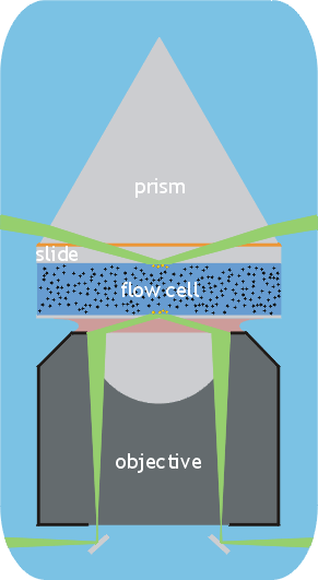
This is probably the easiest configuration to set up, but requires an inverted microscope. A prism is used to couple the incoming laser beam into the microscope slide (see figure). Total internal reflection then occurs at the surface of the microsope slide. The reflected ray is then removed from the slide via the prism. In this case, the main advantage of the prism is that the beam enters and leaves the glass or quartz at a nearly normal incidence. This minimises the intensity of the reflected ray at this interface. A modification of this technique uses two prisms and "bounces" the beam along inside the slide. TIR occurs at multiple points along both surfaces of the slide. This method has the advantage that the area opposite the objective is kept clear and this permits the use of a condenser or other microscope components.
The advantages of this method are that it does not require a special high NA objective (1.4 or greater), although a high NA is still desirable to maximise the collection of light, especially when dealing with intrinsically faint objects such as single molecules. Furthermore, because the excitation beam does not enter the objective lens, scattering and reflections do not occur, minimising the amount of scattered excitation light that has to be filtered out.
The principal disadvantage of the method is that the evanescent wave is generated on the opposite side of the flow cell to the objective lens. If an oil immersion lens is used, then spherical aberration becomes a problem once the thickness of the flow cell exceeds a few tens of microns. Water immersion lenses work better with thicker flow cells, but their overall performance is poorer (see Conibear and Bagshaw (2000)). Thus the method is not as suitable for thicker objects such as cells. Also, the additional manipulations of adding and removing the prism, and realigning the beam, which are necessary every time a new slide is used, make this method more laborious than the objective method.
Typically the laser beam will be brought to a focus at the point where the evanescent wave is generated. This generates an elliptical TIR "footprint" several tens of microns across. In practical use it is often desirable to change the angle of incidence, and to ensure that all the incident light enters at an angle greater than the critical angle. It is therefore desirable that the angle of convergence of the beam is as small as possible, in other words a long focal length lens must be used. Translating the laser beam across this lens will result in a rotation of the beam about the focal point; in other words the angle of incidence can be adjusted without moving the spot. A typical set-up would also include one or two mirrors to steer and align the beam.
To reduce levels of background excitation light, and for laser safety, it is desirable that the output beam be terminated with some kind of "beam dump". Commercial beam dumps are available, but an effective device can be constructed for very little money from a stack of razor blades!
This form of TIRF is ideal for cell biological applications, because the evanescent wave is generated at the cover slip surface and hence the specimen thickness is not an issue. It is particularly useful for investigating processes that occur at or near the cell surface, such as attachment, protrusion or secretion.
In this configuration (see figure) the laser beam is introduced into the periphery of the back aperture of the objective. By focussing the laser beam at the back focal plane of the objective, a parallel beam emerges from the objective lens. The angle at which the beam emerges from the objective depends on the position in the back focal plane where it enters. As the beam moves towards the periphery of the back focal plane, the angle at which the rays emerge increases, until, at the critical angle, TIR occurs. If the optics are set up correctly, the reflected beam should re-enter the objective and return to a focus at the back focal plane.
This technique has several advantages; apart from being ideal for looking at cells, or other thick specimens, it is much less laborious to use, since slides can just be exchanged as in normal microscopy. However, the task of adapting a normal microscope is much more difficult. Another advantage is the completely free access to the specimen from the other side - making it compatible with a very wide range of techniques.
A high NA objective lens is required for this technique - at least 1.4. Many standard objective lenses which are labelled as 1.4 are in fact "rounded up" from their actual values of 1.36 or similar. To achieve TIR, the excitation light must be introduced into the objective at an NA greater than 1.33 (for an aqueous specimen - higher for a typical cell). A finite width is needed to introduce the beam, particularly if it is desired to use angles greater than the critical angle. Some manufacturers now make specialist objectives for TIRF, with NAs as high as 1.65. However, to achieve these exceptionally high NAs, special cover slips and immersion media must be used.
To bring the laser beam into, and out of, the objective, small mirrors can be used, mounted directly below the objective. They should be positioned so as to minimally occlude the light passing through the objective. As with the prism method, the outgoing beam should be removed from the microscope as cleanly as possible to minimise scattered excitation light. Since the light is focussed at the back aperture of the objective, the size of the illuminated area can be controlled by changing the cone angle of the conversing rays. This is achieved by expanding the beam using a Galilean beam expander and then using an adjustable iris diaphragm to control the beam diameter. The beam is then focussed at the back aperture with another lens.
| Contents | Next |