| |
Contents | EBJ Home | Single molecule measurements and biological motors |
 |
||
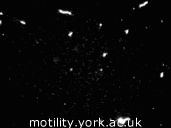
| An in vitro motility assay with rabbit actin and HMM. | ||
The actomyosin motility assay, originally developed in 1986 (see Scholey, 1993), was a key technical development on the way to making single molecule measurements from molecular motors. Furthermore, it shares many practical aspects and reagents with single molecule techniques. Indeed, a common prelude to a single molecule experiment with a new myosin preparation is to test it with a motility assay. The motility assay itself enabled many discoveries in the motors field, particularly the characterisation of the motility of “non-muscle” myosins.
Motility assays have many variations. Typically, however, fluorescently labelled actin filaments slide over a microscope cover slip coated with myosin in the presence of ATP. A sensitive video camera is used to record the motility, and the filaments are tracked by computer, allowing the speed of sliding to be determined. For example, rabbit skeletal muscle myosin and actin produce a sliding velocity of around 5-9 μm·s-1, similar to the maximum shortening velocity of intact muscle.
These assays are very powerful, because the motile system is reconstituted from its components under controlled conditions. This gives the experimenter great control and flexibility in experimental design. For example, different proteolytic fragments of myosin were used in the motility assay to establish that myosin subfragment-1 (containing the motor and regulatory domains) is sufficient for motility.
A typical motility assay involves the following stages:
The motility observed in an in vitro assay is unloaded, in other words the myosin is not acting against a force (the viscous drag on an actin filament moving lengthwise is very small). This means that the motor is able to produce its maximum velocity, but to really characterise the motor we would like to know also about the force it can produce, and the relationship between force and velocity. Furthermore, we would like to be able to determine the contribution made by the individual myosin molecules. To address this, we need to combine the biochemistry of the motility assay with the technique of optical tweezers.
| Contents | Next |